
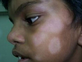
Pityriasis Alba : This disease manifests as ill-defined, hypopigmented macules of varying sizes occurring chiefly on the face of children. This disease is more common in the colder climates. The lesions are asymptomatic and keep on lisappearing spontancously and reappearing till the child atains adulthood. It does not occur in the adults
It is generally believed to be caused by saprophytic staphylococci.
Pityriasis Rosea : This disease is common in the young adults and manifests as a sudden eruption of asymptomatic, 1-2 cm oval or circular, annular lesions which have a pinkish periphery and a scaly centre. The eruption is characteristically localized to the upper trunk and the arms, but may also be restricted to the extremities or the neck. Involvement of the face is rare. The eruption tends to subside spontaneously within 4-6 weeks without leaving any
marks. One attack leads to a life-long immunity. Second attacks are rare. This disease behaves like an exanthem but the causative agent has not so far been established.
Lichen Simplex Chronicus : This disease manifests as a localized area of severely itchy and thickened skin with pigmentation and increased skim markings Such a lesion may be located on any part of the body, but most commonly it occurs on the back of the neck, the leg or the ankle Exacerbations in the lesion are often associated with psychologic stress. Sometimes persistentitching over the lesions of another skin disease such astinea corporis,.psoriasis, lichen planus,contact dermatitis atopic dermatitis, insect bite or infectious eczematoid dermatitis can lead to thickening of the skin which is called secondary lichen simplex chronicus or secondary lichenitication.
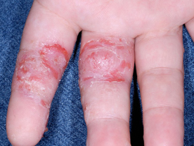
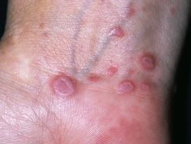
Pompholyx :This term is used for a sudden eruption of severely itchy deep-seated vesicles appearing on the palms and/or sole The fingers and the toes are also usually involved. The are evenly distributed and bilaterally The lesions scale off within a few days but may recur at variable intervals This disease is believed to be caused by an allergic response to an infection somewhere in the body, or a food or an inhalant, but in most cases the cause remains unknown.
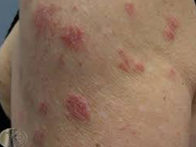
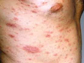
Nummular Eczema : In this disease, the eruptions consist of circular, coin-shaped areas composed of papulo-vesicles, exudation and crusting. The lesions are bilaterally symmetrical and especially located on the extensor aspects of the upper and ower extremities. The eruptions are usually recurrent it is believed to be based on an allergic mechanisnm but the cause of this reaction remains untraced in most of the patients.
Lichen Planus : This disease can manifest in a varicty of clinical forms The most common variant manifests as severely itchy violaceous-pink papules with a smooth shiny surface or mild scaling. Such lesions may be situated on any part of the body though the face is generally not involved. The lesions persist for prolonged periods, but leave behind deeply hyperpigmented macular areas. New lesions may keep appearing as the old ones are subsiding. Several cases in addition have whitish papules or criss-cross streaks on the mucous membranes of the mouth and/or the genitals Some patients also develop thinning of the nail plate which may progress to complete loss of the nail plate (anychia) and formation of pterygium.
Lichen Planus Hypertrophicus : It is a variant of lichen planus in which the lesions are large and hyperkeratotic. Such lesions are commonly located on the legs.
Lichen Plano-Pilaris : It is another variant where the lesions appear at the openings of the hair follicles, and are therefore commonly located on the forearms and legs. Involvement of the scalp leads to cicatricial alopecia.
Actinic Lichen Planus : It is aggravated by sunlight, and therefore the lesions are predominantly located on the sun-exposed areas The extensor surfaces of the forearms are commonly involved and the lesions are usually annular.
Linear Lichen Planus : Lichen planus lesions may also be distributed in a linear fashion on the extremities. Rarely, the lesions may have a segmental distribution when it is called segmental lichen planus. Such lesions are usually located on the trunk. Lichen planus or lichen planus-like lesions can also be precipitated by certain drugs or delayed type graft-versus-host reaction. The cause of lichen planus is not known, though it is believed to be an immunological reaction to an epidermal antigen.
Diagnosis : The diagnosis can he contirmed by histopathology which is characteristic.
Psoriasis : This disease also manifests in a variety of clinical forms.
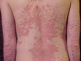
Plaque Type Psoriasis : This is the most frequent form in which the lesions consist of very well-defined erythematous and scaly plaques. The scales are characteristically loosely adherent and become silvery when an attempt is made to scrape them off the lesion. The shapes and the sizes of the lesions vary from one centimeter to very large plaques. The lesions can appear on any area of the body but the extensor aspects of the elbows and the knees, the trunk and the scalp are commonly involved. Face is usually spared. ltching is variable. Most patients experience spontaneous remissions during the summer and relapses during the winter. A few patients may have involvement of the nails in the form of
multiple pin-point pits in the nail plate, onycholysis on onychodystrophy. A few patients may have involvement of the joints which clinically resembles rheumatoid arthritis. Involvement of the distal interphalangeal joints however, is characteristic of psoriasis arthropathy. The hain and the mucous membranes are not involved.
Psoriasis Erythroderma : In some patients, the psoriasis may extend to involve the entire body surface and present as generalized redness and scaling all over the body with chills and rigors.
Guttate Psoriasis : It is another variant in which the lesions consist of sudden eruptions of small erythematous and scaly papules or plaques. Such lesions disappear within a few wecks but recurrences are common. Sometimes such lesions may ultimately persist and increase in size to form the plaque type of psoriasis.
Pustular PsoriasisIt is a severe form of the disease in which thc lesions consist of small superficial pustules which may appear on a plaque of psoriasis or oceur independently.
Palmo-Plantar Psoriasis : In some cases, psoriasis involves only the palms and/or the soles. This type is called palmo-plantar psoriasis Occasionally the nails may be the only tissue involved.
Aetiopathogenesis : Plaque type psoriasis is based on a genetic predisposition in which the mechanism which controls the epidermal cell turn-over is defective. In a psoriatic lesion the cells proliferate at a rate ten times faster than the normal cpidermis. Trauma plays a significant role in precipitating the lesions. Some patients display a tendency for the Koebner phenomenon. Psoriasis erythroderma can result from irritant topical therapy for plaque psoriasis. Guttate psoriasis is believed to be an immune complex
disease precipitated by streptococcal throat infection or s bacteria. Pustular psoriasis indicates a high degree of inflammation where the Munro's micro-abscesses become large
enough to be visible clinically.
Diagnosis : The diagnosis of psoriasis can be confirmed histopathologically which shows very distinctive histopathology.
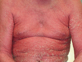
Erythroderma : This disease is also called exfoliative dermatitis. In this disease the skin over the entire body develops erythema and scaling. Because of generalized vasodilatation, the skin loses its ability to control the loss of body heat and therefore develops chills and rigors. In severe cases the hain and the nails may also be shed off, and there is a tendency to develop secondary pyogenic infection of the skin and other organs. Erythroderma can result from a pre-existing skin disease such as psoriasis, atopic dermatitis, seborrhocic dermatitis. air-borne contact dermatitis, pityriasis rubra pilaris and less commonly other dermatoses. Injudicious irritating topical treatment of the primary disease can precipitate this transformation. Erythroderma can also result from an uncontrolled exanthematous drug reaction. Less commonly erythroderma may represent a cutaneous manifestation of internal malignancy.
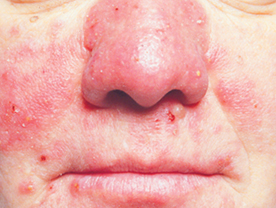
Rosacea : This disease is common in middle-aged ladies and manifests as recurrent episodes of erythema on the face, especially the cheeks. Gradually the erythema becomes persistent and the patient may also develop telangiectasia and erythematous accuminate papules.
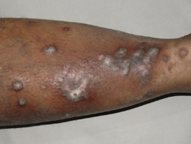
Prurigo Nodularis : This disease manifests as multiple hyperpigmented nodules on the legs, forearms and occasionally on other parts of the body. The lesions are extremely itchy and often excoriated. Some of the nodules may be partly depigmented. The cause of this disease is not known, the possibility of an exaggerated response to insect bites is worth considering
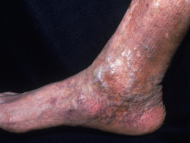
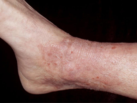
Stasis Dermatitis : Patients having varicose veins are likely to develop telangiectasia, pigmented spots and even ulcers on the ankle, lower part of the leg and the foot. These ulcers can become chronic if the patient does not take adequate care.